SLO-Net
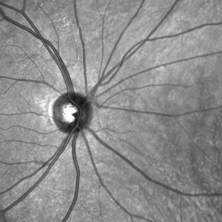
Purpose: Several machine learning studies have used optical coherence tomography (OCT) for multiple sclerosis (MS) classification with promising outcomes. Infrared reflectance scanning laser ophthalmoscopy (IR-SLO) captures high-resolution fundus images, commonly combined with OCT for fixed B-scan positions. However, no machine learning research has utilized IR-SLO images for automated MS diagnosis.
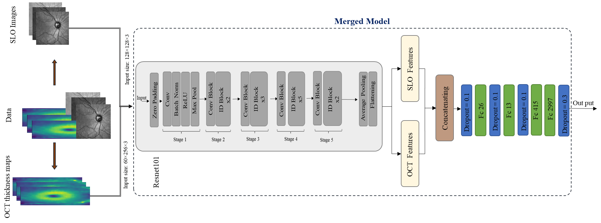
Methods: This study utilized a dataset comprised of IR-SLO images and OCT data from Isfahan, Iran, encompassing 32 MS and 70 healthy individuals. A number of convolutional neural networks (CNNs)—namely, VGG-16, VGG-19, ResNet-50, ResNet-101, and a custom architecture—were trained with both IR-SLO images and OCT thickness maps as two separate input datasets. The highest performing models for each modality were then integrated to create a bimodal model that receives the combination of OCT thickness maps and IR-SLO images. Subject-wise data splitting was employed to prevent data leakage among training, validation, and testing sets.
Results: Overall, images of the 102 patients from the internal dataset were divided into test, validation, and training subsets. Subsequently, we employed a bootstrapping approach on the training data through iterative sampling with replacement. The performance of the proposed bimodal model was evaluated on the internal test dataset, demonstrating an accuracy of 92.40% ± 4.1% (95% confidence interval [CI], 83.61–98.08), sensitivity of 95.43% ± 5.75% (95% CI, 83.71–100.0), specificity of 92.82% ± 3.72% (95% CI, 81.15–96.77), area under the receiver operating characteristic (AUROC) curve of 96.99% ± 2.99% (95% CI, 86.11–99.78), and area under the precision–recall curve (AUPRC) of 97.27% ± 2.94% (95% CI, 86.83–99.83). Furthermore, to assess the model generalization ability, we examined its performance on an external test dataset following the same bootstrap methodology, achieving promising results, with accuracy of 85.43% ± 0.08% (95% CI, 71.43–100.0), sensitivity of 97.33% ± 0.06% (95% CI, 83.33–100.0), specificity of 84.6% ± 0.10% (95% CI, 71.43–100.0), AUROC curve of 99.67% ± 0.02% (95% CI, 95.63–100.0), and AUPRC of 99.65% ± 0.02% (95% CI, 94.90–100.0).
Conclusions: Incorporating both modalities improves the performance of automated diagnosis of MS, showcasing the potential of utilizing IR-SLO as a complementary tool alongside OCT.
Translational Relevance: Should the results of our proposed bimodal model be validated in future work with larger and more diverse datasets, diagnosis of MS based on both OCT and IR-SLO can be reliably integrated into routine clinical practice.